
Prevent, Stabilize, Reverse Macular Degeneration
Welcome to the Macular Program
The Macular Program is specifically tailored for individuals affected by macular degeneration. This condition can stem from various factors, including hormonal decline, oxidative stress, inflammation, and general disruptions in the body's chemical processes. The goal of our program is to rejuvenate the cells in the macula to improve their function and halt further deterioration.
Our approach is centered on a personalized, multi-modal strategy that factors in your medical history and biochemical profile derived from comprehensive laboratory assessments. Your individualized care plan will feature specific nutraceuticals and, where necessary, bio-identical hormones. These interventions are designed to optimize your cellular health and reinvigorate the functionality of your genes.
To halt the progression of macular degeneration most effectively, it's crucial to initiate treatment as soon as possible, before macula damage becomes permanent. Once the damage is irreversible, our options are limited to stabilizing the condition. However, early intervention can potentially reverse vision loss.
The Macular Program is specifically tailored for individuals affected by macular degeneration. This condition can stem from various factors, including hormonal decline, oxidative stress, inflammation, and general disruptions in the body's chemical processes. The goal of our program is to rejuvenate the cells in the macula to improve their function and halt further deterioration.
Our approach includes a targeted mix of hormonal treatments, carefully chosen nutraceuticals, and personalized nutritional guidance derived from individual lab tests. We aim to reactivate genes that have become inactive as a result of aging. Our studies indicate that this method can not only stabilize but potentially enhance vision in nearly half of the participants suffering from macular degeneration. Additionally, it could decrease the frequency of eye injections and reduce the chances of the condition worsening from the dry type to the more severe wet type.
We highly advocate for patients to undertake this program, which is grounded in scientific research, as soon as possible. Early participation can significantly diminish the likelihood of the disease advancing to an irreversible stage.
Welcome to the Macular Program
We highly recommend patients to become part of this research-backed program as soon as possible. Participating early can substantially mitigate the risk of the disease becoming irreversible.
To discover how this may benefit you, click the button 👇 below to Speak to a Specialist.
After viewing the video, click the button 👇 to “Book a Call” or Speak to an Advisor.

Prevent, Stabilize, Reverse Macular Degeneration
⬇️ Watch The Educational Video And Book Your Free Consultation ⬇️
Welcome to the Macular Program
The Macular Program is specifically tailored for individuals affected by macular degeneration. This condition can stem from various factors, including hormonal decline, oxidative stress, inflammation, and general disruptions in the body's chemical processes. The goal of our program is to rejuvenate the cells in the macula to improve their function and halt further deterioration.
Our approach is centered on a personalized, multi-modal strategy that factors in your medical history and biochemical profile derived from comprehensive laboratory assessments. Your individualized care plan will feature specific nutraceuticals and, where necessary, bio-identical hormones. These interventions are designed to optimize your cellular health and reinvigorate the functionality of your genes.
To halt the progression of macular degeneration most effectively, it's crucial to initiate treatment as soon as possible, before macula damage becomes permanent. Once the damage is irreversible, our options are limited to stabilizing the condition. However, early intervention can potentially reverse vision loss.
We highly recommend patients to become part of this research-backed program as soon as possible. Participating early can substantially mitigate the risk of the disease becoming irreversible.
To discover how this may benefit you, click the button 👇 below to Speak to a Specialist.

Symptoms of the disease
Difficulty seeing in dim light.
Slowly progressive blurred vision not improved with glasses.
Distorted Vision: Straight lines might appear wavy or curved, which is a sign of wet macular degeneration.
Spots: There may be dark blind spots in the center of vision which may be transitory.

Macular Degeneration symptoms usually develop gradually and without pain. Over time, as the condition worsens, symptoms become more pronounced and can significantly affect daily activities like reading, driving and recognizing faces.
Symptoms of the disease

Difficulty seeing in dim light..
Slowly progressive blurred vision not improved with glasses.
Distorted Vision: Straight lines might appear wavy or curved, which is a sign of wet macular degeneration.
Spots: There may be dark blind spots in the center of vision which may be transitory
Macular Degeneration symptoms usually develop gradually and without pain. Over time, as the condition worsens, symptoms become more pronounced and can significantly affect daily activities like reading, driving and recognizing faces.


Our Methodology
Our team of medical experts develop custom-tailored care plans involving nutrition, nutraceuticals and hormones to optimize cellular biochemistry. These care plans are based on history and blood testing and enhance eye and overall health and well being. The goal is to restore optimal cellular function to stop or reverse the “degeneration” of cells.
Introducing the HOMING Method to Combat
Macular Degeneration

Our Methodology

Our team of medical experts develop custom-tailored care plans involving nutrition, nutraceuticals and hormones to optimize cellular biochemistry. These care plans are based on history and blood testing and enhance eye and overall health and well being. The goal is to restore optimal cellular function to stop or reverse the “degeneration” of cells.
The HOMING Method
The HOMING Method
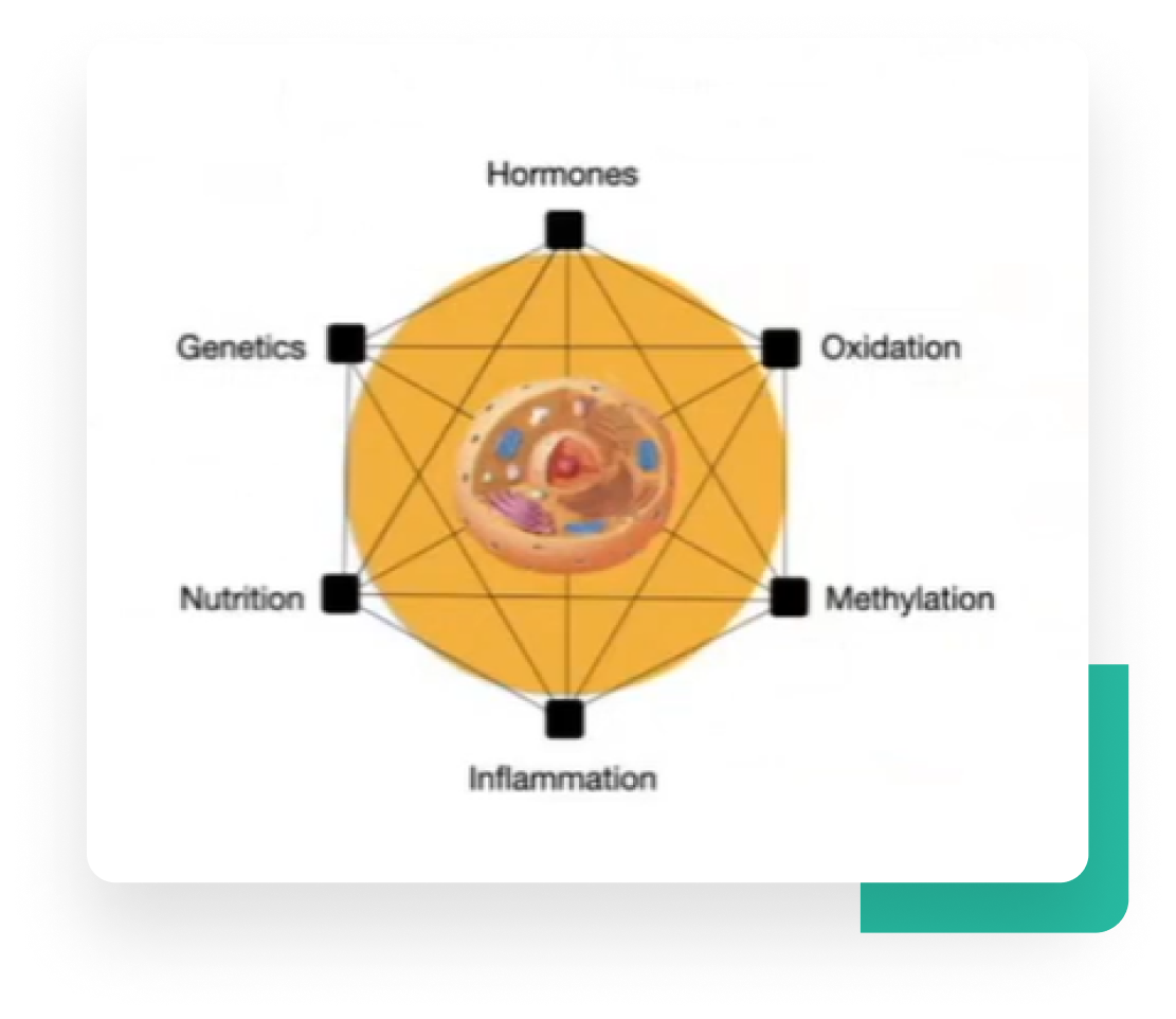
The HOMING Method guides the personalized care plan for Age-related Macular Degeneration (AMD).
HOMING is an acronym for Hormones, Oxidation, Methylation, Inflammation, Nutrition and Genetics. The goal is to improve cell biochemistry either directly or by epigenetic mechanisms, (reawakening dormant genes that down regulate with aging)
Literature Citation demonstrating stability and improvement of dry and wet AMD
Let’s Talk about Your
Dry or Wet AMD Goals
Let’s Talk about Your
Dry or Wet AMD Goals


In 2020, we conducted a study with 365 dry or wet AMD patients. We found that stability or improvement was possible for dry or wet AMD.
48% of dry AMD improved.
60% of wet AMD improved.

In 2020, we conducted a study with 365 dry or wet AMD patients. We found that stability or improvement was possible for dry or wet AMD.
It won the Advancements in Health Care Award.
Had it not been for our success in many patients we may never have pursued macular degeneration.
We discuss the discovery process in our presentations.
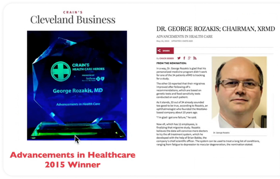
The HOMING method has been a success in many clinical conditions indicating that addresses root causes of disease.
It won the Advancements in Health Care Award.
Had it not been for our success in many patients we may never have pursued macular degeneration.
We discuss the discovery process in our presentations.

Who will be eligible for
the Program

Prevent AMD
Failures of Dark Adaptation Testing
Drusen
Dry AMD with Vision Loss
Reducing conversion of Dry to Wet AMD
Wet AMD
Diabetic Retinopathy
Vein Occlusions
Preparing for Cataract Surgery in Eyes with AMD
Glaucoma
Retinitis Pigmentosa

Macular hole
Artery Occlusion
Macular Pucker (Epiretinal Membrane)
Floaters
Retinal Detachment
Irreversible Advance Macular Degeneration

Who will be eligible for
the Program

Prevent AMD
Failures of Dark Adaptation Testing
Drusen
Dry AMD with Vision Loss
Reducing conversion of Dry to Wet AMD
Wet AMD
Diabetic Retinopathy
Vein Occlusions
Preparing for Cataract Surgery in Eyes with AMD
Glaucoma
Retinitis Pigmentosa

Macular hole
Artery Occlusion
Macular Pucker (Epiretinal Membrane)
Floaters
Retinal Detachment
Irreversible Advance Macular Degeneration
The Experts behind the Program

George W. Rozakis MD
Board Certified Ophthalmology

Brian A. Bakke, Ph.D
Ph.D. in Biochemistry
The Experts behind the Program

George W. Rozakis, MD
Board Certified Ophthalmologist / Biomedical Engineer

Brian Bakke, Ph.D
PhD in Biochemistry

What our patients say

Calvin K.
I am extremely pleased with the program. My eyesight has improved, especially my long distance vision for driving. I am able to drive at night!

What our patients say

Calvin K.
I am extremely pleased with the program. My eyesight has improved, especially my long distance vision for driving. I am able to drive at night!

Macular Program Articles
Unlock the Secrets of Drusen: Key Insights into Preventing Macular Degeneration
“Stabilizing And Reversing Macular Degeneration With A Cellular Medicine Approach Is The Key To Protecting Your Vision and Managing General Health Concerns .” - The Macular Program
Unlock the Secrets of Drusen: Key Insights into Preventing Macular Degeneration
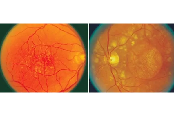
Macular degeneration, a leading cause of vision loss, often begins with the appearance of drusen. These tiny yellow or white deposits under the retina are more than just an eye anomaly; they are the early warning signs of potential retinal damage.
What Are Drusen?
Drusen are small lipid-rich deposits that form beneath the retina. They are often detected during routine eye examinations and can vary in size. While they are commonly found in older adults, their presence in larger sizes or in greater quantities can indicate the early stages of Age-related Macular Degeneration (AMD).
Visual Evidence: The Importance of Pictures and OCT Scans
To accurately diagnose and monitor the progression of macular degeneration, eye care professionals rely heavily on retinal imaging. Two key methods include:
1. Fundus Photography
This captures detailed images of the retina, highlighting the presence of drusen. These photos provide a clear, color view of drusen, appearing as small yellowish spots on the retina.
2. Optical Coherence Tomography (OCT)
OCT is a more advanced technique, offering a cross-sectional view of the retina. It reveals the size, shape, and layering of drusen, providing crucial information about the health of the macula and the risk of progression to AMD.
The pathogenesis of drusen, particularly in the context of age-related macular degeneration (AMD), is a complex process involving multiple factors and pathways. Here's an integrated overview:
Metabolic Dysfunction in the Retina: The retinal pigment epithelium (RPE) cells, which are crucial for the health of the retina, become less efficient at processing and clearing metabolic waste products as we age. This inefficiency leads to the accumulation of cellular debris, including lipids, proteins, and other materials, forming drusen beneath the RPE.
Lipid Accumulation and Cholesterol: Drusen contain significant amounts of lipids, including cholesterol. Dysregulation in lipid metabolism, either locally in the eye or as part of systemic conditions like hypercholesterolemia, contributes to the accumulation of cholesterol in drusen.
Inflammatory Response: Chronic low-grade inflammation is a key factor in the formation of drusen. Inflammatory cells are recruited to the retina in response to cellular stress, damage, or the presence of abnormal deposits. These cells, along with the release of inflammatory mediators, contribute to the development and growth of drusen.
Oxidative Stress: Oxidative damage to retinal cells, induced by factors such as UV light exposure and smoking, leads to the production of reactive oxygen species (ROS). ROS can damage cellular components, contributing to the accumulation of waste products that form drusen.
Immune System Involvement: Components of the immune system, particularly the complement system, are involved in the pathogenesis of drusen. Dysregulation of the complement system can lead to chronic inflammation and tissue damage in the retina, promoting drusen formation.
Blood-Retina Barrier (BRB) Dysfunction: A compromised BRB can allow the infiltration of blood-derived substances and inflammatory cells into the retinal tissue, further contributing to the formation of drusen.
Genetic Factors: Certain genetic variants, particularly in genes related to the complement pathway and lipid metabolism, have been associated with an increased risk of AMD and drusen formation.
Environmental and Lifestyle Factors: Environmental factors like smoking, diet, and sunlight exposure, along with lifestyle choices, influence the risk of drusen formation. For instance, smoking is a known risk factor for AMD and can exacerbate oxidative stress and inflammation in the retina.
In summary, the pathogenesis of drusen is multifactorial, involving age-related metabolic changes, lipid accumulation, chronic inflammation, oxidative stress, immune system dysregulation, genetic predispositions, and environmental influences. This complex interplay leads to the accumulation of extracellular material beneath the RPE, forming the characteristic deposits known as drusen. Understanding these pathways is crucial for developing prevention and treatment strategies for AMD and other related retinal diseases.
Drusen Explained: The Link to Macular Degeneration
Drusen themselves do not usually cause vision loss. However, their presence, especially in larger sizes or in significant numbers, is a key indicator of AMD.
This is often associated with the presence of many medium-sized drusen. As these drusen grow and become more numerous, they can lead to a thinning and drying out of the macula, leading to gradual vision loss.
Conclusion
Regular eye exams that include the screening for drusen are vital, especially for individuals over the age of 50. Early detection of drusen can lead to proactive monitoring and management of eye health, potentially slowing the progression of AMD and preserving vision.
Remember, knowledge about drusen is a crucial step in understanding and combating macular degeneration. Keep your eyes on the prize – maintaining healthy vision – by staying informed and vigilant.
The Macular Program manages many of these underlying causes of drusen. In our clinical experience, drusen either stop progressing or improve. Unfortunately, drusen tends to be irreversible, so it's very important to arrest their progression as soon as possible.
Need to stop progressive vision loss?
Register for the next LIVE Macular Masterclass here.
Patients FAQ'S
Got Questions? We’ve Got Answers.
How long does it take to see improvements with dry AMD?
Our study followed patients for 6 months. Those who improved did so generally between 2 and 6 months. Unpublished data is showing that these effects are not lost out to 2-3 years and additional improvement is possible with more time. Time and compliance with the program are the keys to continued success.
Do I need to continue to take the program to sustain the benefits?
Similar to exercise and dietary practices, the favorable changes in your blood chemistry can only be sustained as long as you are taking your program.
Do Blood Tests Improve?
Yes. Consistently adhering to our program has led to marked improvements for our patients. It is the combination of time and unwavering commitment to the program that paves the way for enduring success.
How do you decrease the frequency of injections for Wet AMD.
This decision is made by you retina doctor. However it is reasonable to ask to decrease the time between injection if the OCT scans show no remaining fluid because the HOMING Method, according to scientific literature, may decrease the creation of growth factors in the eye (for which you are receiving the injections).
Is this Anti-Aging Medicine?
The Macular Program is a form of anti-aging medicine since macular degeneration is actually called AGE RELATED macular degeneration. Fighting aging is fighting AMD. What is nice is that treating AMD with HOMING also benefits other aging symptoms which is why we want to know about your eye AND General Health.
What would slow improvement in Dry or Wet AMD?
Age-Related Macular Degeneration (AMD), whether dry or wet, is a complex condition influenced by numerous factors. Our HOMING method helps to prevent, stabilize and even reverse macular degeneration.
What about GA?
Geographic Atrophy is a special form of dry macular degeneration that is felt to be caused by inflammation. The HOMING method reduces inflammation. Our clinical suspicion is that GA does not worsen if a patient is on the HOMING Method. This is an important matter because new drugs now exist to fight inflammation in eyes with GA, but these drugs need to be injected into the eye. We hope the HOMING method will allow patients to avoid eye injections for GA. The smart move is to be on the HOMING method and have the GA closely followed for progression. If no progression, then you don’t need shots. It’s your call.


New breakthrough allows age-related macular degeneration patients to improve vision and prevent further loss.
MacularProgram© 2023 | All Rights Reserved
All Rights Reserved 2024
Macular Program
Contact Us:
440-777-2667
Email Us: info@macularprogram.com

